Unknown Insect Genus Trapped In Amber For Over 35 Million Years – New Discovery
Eddie Gonzales Jr. – MessageToEagle.com – Thanks to an international research collaboration involving the University of Granada (UGR), a hitherto undescribed species of insect has been discovered: Calliarcys antiquus, which belongs to the Ephemeroptera (mayfly) order.
The specimen itself was located by Arnold Staniczek of Stuttgart’s State Museum of Natural History, set in a piece of Baltic amber estimated to be between 35 and 47 million years old.
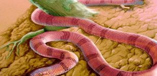
Image of the insect. Credit: University of Granada
And, thanks to the specialist contribution of Professor Javier Alba-Tercedor of the UGR’s Department of Zoology, using microtomography to obtain clear images of the insect, it could be studied and described in detail.
Plants such as conifers (and certain legumes) protect themselves by secreting resin—a thick, sticky liquid—as a reaction to damage to the cortex of the specimen. As insects often become trapped in this resin, even those dating back millions of years may still be found to this day, preserved in the hardened, fossilized resin that we know as amber. There are amber deposits located in different parts of the world, including northern Spain, but those located in the Baltic region are the most abundant.
“The conservation of the specimens trapped inside the amber is often excellent, and the transparency of the material that surrounds them enables them to be studied, under a microscope, in great detail,” explains Professor Alba-Tercedor.
“But, in other cases, the level of transparency is not good because the areas of opacity that form prevent certain details from being examined,” comments Alba-Tercedor. When this limited transparency is problematic, X-ray microtomography (a technique similar to that used in hospitals to study patients’ organs) is invaluable in studying fossil specimens that are preserved in amber.
When Arnold Staniczek—a specialist in Ephemeroptera, with extensive experience in the study of insects preserved in amber—observed this particular piece from the Baltic, it was completely transparent. However, the insect itself presented certain “hyaline” (translucent) areas surrounding certain parts of the body that are essential for characterizing the specimen and distinguishing one species from another, such as the end of the abdomen where the male reproductive apparatus (genitalia) are located.
As this translucence impeded the identification process, Staniczek turned to Alba-Tercedor, in his capacity as a specialist in Ephemeroptera and due to his recognized experience in the use of computerized microtomography (micro-CT) applied to the study of insects.
From his base at the microtomography unit of the UGR’s Department of Zoology, Professor Alba-Tercedor reconstructed the entire insect, including those areas otherwise impossible to observe due to the opacity of the amber. This particular specimen belongs to the genus Calliarcys, the first (formally described) species of which is found in the Iberian Peninsula.
Thanks to the expert knowledge of Roman Godunko of the Institute of Entomology of the Czech Academy of Sciences, the study of the previously undescribed species of mayfly was then accomplished by comparing it with extant species of the genus. In addition, given the importance of molecular studies in characterizing species and determining their evolutionary position, the input of Polish experts from the University of Lódz was also sought.
Hence, researchers Michal Grabowski and Tomasz Rewicz completed the study with a DNA analysis of extant species of the genus.
“In short, it all started with the discovery of a beautiful insect preserved in amber, which attracted the attention of the expert eyes of a scientist. And which ultimately required the enthusiastic collaboration and detective work of five scientists based in research centers located in four countries, who, after applying the latest techniques, were finally able to name and describe an insect that has remained locked inside a drop of amber for millions of years,” says Professor Alba-Tercedor.
What is micro-CT?
Micro-CT is a technique for producing a 3D image using X-rays. It uses the same method as computed tomography (CT) in medicine, but on a smaller scale and with a much higher resolution. While CT provides a resolution measured in millimeters, in micro-CT, resolutions of around 0.5 micrometers are achieved.
Indeed, the new nano-CTs are increasing the resolution even further and expanding the possibilities the technology can offer. Micro-CT is based on 3D microscopy, which enables the internal structure of extremely small-scale objects to be captured non-invasively.
No physical cross-sections or complex pre-treatments are required: with just a single scan, multiple radiographic images can be generated to obtain high-resolution 3D rendered images of the entire internal structure of the sample. And all of this while the sample is left intact for subsequent treatments.
In simple terms, the procedure consists of an X-ray source illuminating the object and a flat X-ray detector capturing enlarged projection-images. Using computer software, the X-rays derived from the sample are then transformed into cross-sections that are converted into three-dimensional images using volumetric reconstruction programs.
The findings were published in Scientific Reports.
Written by Eddie Gonzales Jr. – MessageToEagle.com Staff


